
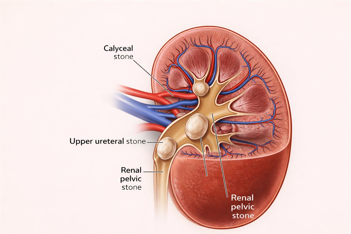
Renal Stones (Kidney Stones)
Renal stones are hard mineral deposits that form inside the kidneys due to crystallization of substances present in urine.
These stones may remain in the kidney or move into the ureter, causing pain, obstruction, and infection.
Renal stones are common, recurrent, and largely preventable with proper treatment and lifestyle measures.
How Do Renal Stones Form
When urine becomes concentrated due to low fluid intake or metabolic imbalance, minerals such as calcium, oxalate, uric acid, or cystine crystallize.
Over time, these crystals aggregate and form stones. Dehydration, dietary habits, infections, and metabolic disorders play a major role.
Common Symptoms
- Sudden severe pain in the flank or lower back
- Pain radiating to the lower abdomen or groin
- Nausea and vomiting
- Burning sensation during urination
- Blood in urine
- Frequent urge to pass urine
- Some small stones may remain asymptomatic
When to Seek Medical Help
- Pain that is severe, persistent, or not relieved with medication
- Blood in urine that continues or increases
- Fever, chills, or signs of infection
- Difficulty or inability to pass urine
- Pain associated with repeated vomiting
- Known kidney disease or single kidney with pain symptoms
- Recurrent stone episodes or worsening symptoms
- Symptoms interfering with daily activities or work
Early medical evaluation helps prevent infection, obstruction, sepsis, and permanent kidney damage.
Types of Renal Stones
Calcium Stones
Most common type
- Composed of calcium oxalate or calcium phosphate
- Associated with dehydration, high salt intake, and metabolic imbalance
Uric Acid Stones
Form due to excess uric acid
- Common in gout, obesity, diabetes, and high-protein diets
- Related to acidic urine
Struvite Stones
Associated with recurrent urinary tract infections
- Can grow rapidly and form large staghorn calculi
- More common in women
Cystine Stones
Rare and hereditary
- Due to excessive cystine excretion in urine
- Often recurrent and difficult to treat
Risk Factors
- Low water intake and dehydration
- High salt and animal protein diet
- Family history of kidney stones
- Recurrent urinary tract infections
- Obesity and sedentary lifestyle
- Metabolic disorders such as gout
- Certain medications
How Are Renal Stones Diagnosed
- Ultrasound of kidneys, ureters, and bladder
- Non-contrast CT scan for precise size and location
- X-ray KUB for radio-opaque stones
- Urine examination and culture
- Blood tests to assess kidney function and metabolic causes
Treatment Options for Renal Stones
Conservative (Medical) Management
Suitable for small stones
- High fluid intake to increase urine output
- Pain relief medications
- Drugs to relax the ureter and aid stone passage
- Diet modification based on stone composition
Surgical treatments
ESWL (Extracorporeal Shock Wave Lithotripsy)
Used for kidney stones usually up to 2 cm in size
- External shock waves are focused on the stone to break it into fragments
- No incision or endoscope is required
- Stone fragments pass naturally in urine
Best for selected stones with favourable size, position, and hardness
URSL (Ureteroscopic Laser Lithotripsy)
Used for stones located in the ureter and selected small renal stones
- A thin endoscope is passed through the urinary passage
- Laser energy is used to fragment the stone
- Fragments are removed or allowed to pass naturally
- Temporary ureteric stent may be placed
RIRS (Retrograde Intrarenal Surgery – Laser)
Preferred for renal stones up to 2 cm, especially in difficult locations
- A flexible endoscope is passed through the urethra, bladder, and ureter into the kidney
- Holmium or thulium laser is used to dust or fragment stones inside the kidney
- No incision is made
Ideal for multiple stones, lower calyx stones, or ESWL-resistant stones
PCNL (Percutaneous Nephrolithotomy)
Recommended for stones larger than 2 cm or staghorn calculi
- A small incision is made in the back to access the kidney directly
- Stones are removed using nephroscope and may be fragmented using laser or ultrasonic devices
Provides the highest stone clearance in complex cases
Mini-PCNL / Ultra-Mini PCNL
Used for medium-sized stones to reduce tissue trauma
- Smaller access tract compared to standard PCNL
- Laser is commonly used for stone fragmentation
Associated with faster recovery and less pain
Open or Laparoscopic Stone Surgery
- Rarely required in current practice
- Reserved for very large, complex stones or anatomical abnormalities
- Longer recovery compared to minimally invasive laser-based techniques
Complications if Untreated
- Repeated severe pain episodes
- Recurrent urinary tract infections
- Urinary obstruction and kidney swelling
- Permanent kidney damage
- Life-threatening infection (urosepsis)
Recovery and Aftercare
- Rapid recovery after minimally invasive procedures
- Adequate hydration is essential
- Temporary stent-related discomfort may occur
- Normal activities usually resume within 1–3 weeks depending on treatment
- Follow-up imaging to confirm complete stone clearance
Prevention of Renal Stones
- Maintain adequate daily water intake
- Reduce salt consumption
- Balanced diet with controlled animal protein
- Prompt treatment of urinary infections
- Metabolic evaluation in recurrent stone formers
Outlook
Renal stones are highly treatable with modern minimally invasive techniques.
Early diagnosis, correct procedure selection, and preventive strategies significantly reduce recurrence and protect long-term kidney function.